The Use of Neuroimaging in Pain Medicine

The phenomenon of acute pain serves two main purposes. First, it acts as a warning system to the body that the risk from harm is imminent. Second, it indicates that an injury has already occurred, and a period of rest is required while healing takes place. However, once pain has persisted for more than 3 months, it is termed chronic. At this stage, it can cease to be a symptom and become a disease in its own right. The pathophysiology of chronic pain is not well understood and this hinders the development of new effective therapies, which are currently lacking.
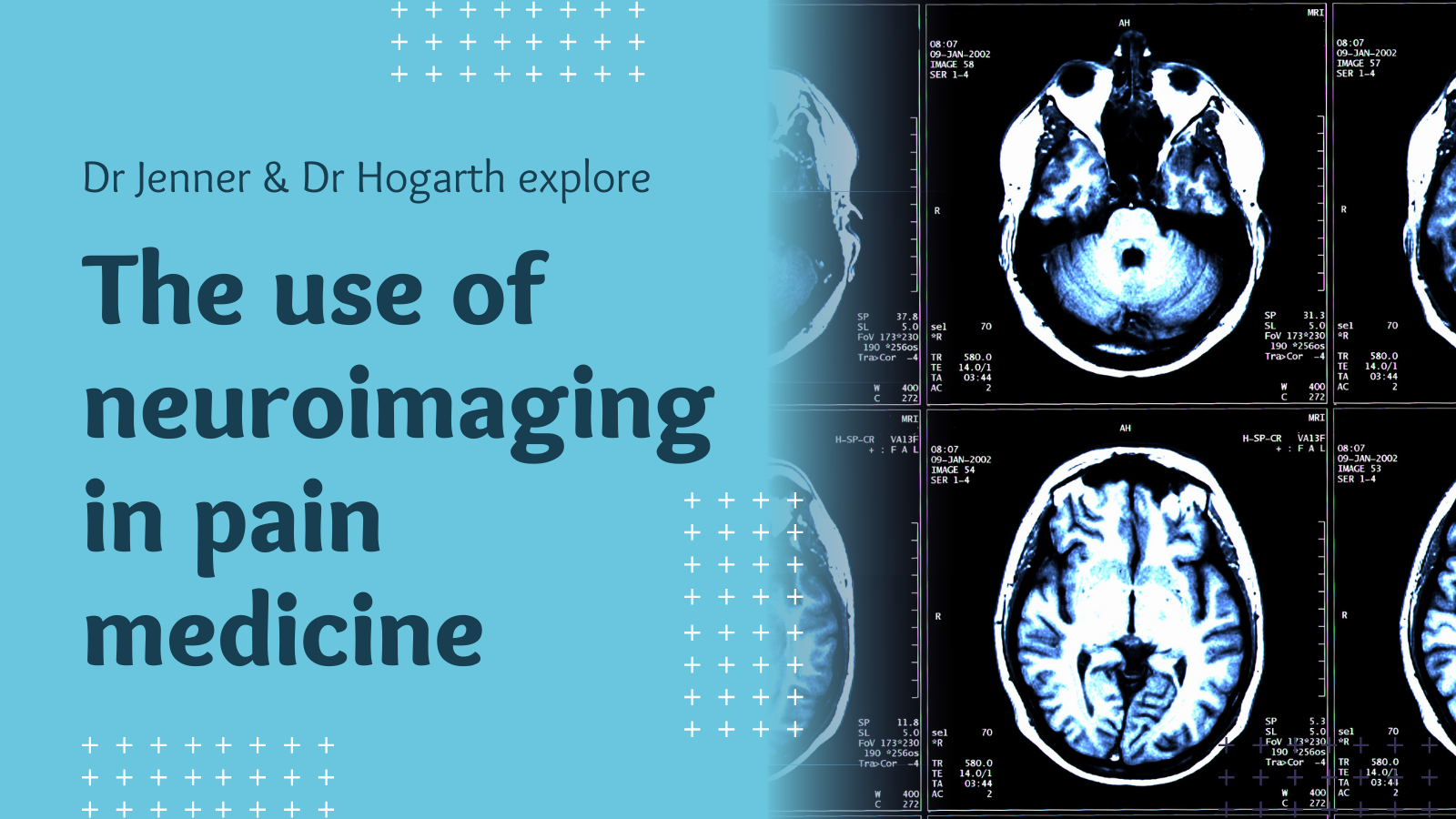
Neuroimaging refers to imaging of the brain, brainstem and spinal cord. The most widely used technique is magnetic resonance imaging (MRI), which uses a strong magnetic field and radiofrequency pulses to produce high resolution images. From these, changes in the brain’s structure and activity can be viewed. Chronic pain is amplified and maintained by complex neural mechanisms, which the current understanding of these processes is limited. However, neuroimaging can provide new insights, and by increasing our understanding, it should be possible to develop new effective treatments for chronic pain.
Pain has both a sensory and an emotional element. Due to differences in the way we perceive these, and variation in cognitive factors, no two people experience pain in exactly the same way. Previously, it has been difficult to study how these factors interact in pain perception, but by identifying related changes in brain activity in key areas responsible for sensory and motor processing, as well as the affective and emotional aspects of pain, neuroimaging will allow their impact to be properly assessed. In chronic pain, it is thought that the normal pathways involved in pain processing become upregulated, which leads to a state known as central sensitisation. This is due to inflammation in the skin causing an increase in nociceptive signals from the peripheral nerves, combined with a lack of inhibition or increased activity within the spinal cord, brainstem or cortex, where pain modulation and central sensitisation occur.
Research has indicated that several structural differences exist in the brains of individuals experiencing chronic pain. Several conditions, including fibromyalgia (FM), complex regional pain syndrome (CRPS), chronic low back pain, migraine and irritable bowel syndrome, have demonstrated changes in cortical thickness and gray matter density. For the latter, these differences usually manifest as a decrease in regional gray matter density. These decreases may be the result of age-related atrophy. However, several studies have reported simultaneous decreases and increases in different regions of the brain, but the primary cause of these changes is unclear. They may be due to pre-existing variation in brain structure, which predisposes the patient to chronic pain. It is also unclear whether these changes play a functional role in the development and maintenance of chronic pain. While they may have occurred as a response to chronic pain, it is also possible that they arise as a result of one of the many comorbid conditions, such as depression, anxiety or sleep disturbance, associated with chronic pain, or through medication use. A meta-analysis of FM studies reported that depression scores accounted for most of the changes in gray matter structure, but similar changes have also been observed in patients with chronic low back pain experiencing very low levels of depression.
Neuroimaging has also indicated that altered brain activity is associated with numerous chronic pain conditions. Rather than there being a single ‘pain centre’, several regions of the brain have consistently demonstrated alterations in function in patients with chronic pain, including regions involved in sensory and motor processing and those dealing with emotions, fear and memory. Examination of the relationships of activity across multiple brain regions, or networks, shows that there are differences in functional connectivity in patients with chronic pain. In particular, the default-mode network (DMN), which is more active when the person is resting, shows greater connectivity with the insular cortex in patients with FM. However, in CRPS, connectivity is decreased within the DMN but increased in pain-processing regions of the brain. Furthermore, negative emotions can augment the perceived pain intensity and increase the pain-related brain activity.
While the majority of neuroimaging studies to date have been cross-sectional, a few longitudinal investigations have been performed. In one, changes in white matter structure followed the transition from subacute to chronic low back pain. Elsewhere, some brain changes have been reversed after treatment. Therefore, it appears that the differences observed in patients with chronic pain may not be permanent.
Neuroimaging has now identified several structural and functional changes within the brains of patients with chronic pain. While this may help to dispel the view that chronic pain is simply a psychological condition, the individual variability in the pain experience remains a challenge for clinicians. Identification of specific regions within the brain that are responsible for the mechanisms that support ongoing chronic pain may provide targets for the development of new therapies. The use of effective therapies may not only reduce the pain but also help to restore normal brain structure and function.
About our experts
Dr Chris Jenner is a Consultant in Pain Medicine at Imperial College NHS Trust. He is a highly experienced expert witness, providing medico legal reports for a wide range of pain conditions for personal injury and medical negligence cases.
Dr Kieran Hogarth is a Consultant Neuroradiologist at the Royal Berkshire Hospital, where he is the lead radiologist for neuroimaging. Dr Hogarth has extensive experience writing medico legal and expert witness reports for both adult and paediatric cases and has attended court on multiple occasions.
Further reading
Martucci, K. T., & Mackey, S. C. (2016). Imaging Pain. Anesthesiology Clinics, 34(2), 255–269. https://doi.org/10.1016/j.anclin.2016.01.001
Wartolowska, K., & Tracey, I. (2009). Neuroimaging as a tool for pain diagnosis and analgesic development. Neurotherapeutics: the Journal of the American Society for Experimental Neurotherapeutics, 6(4), 755–760. https://doi.org/10.1016/j.nurt.2009.08.003


